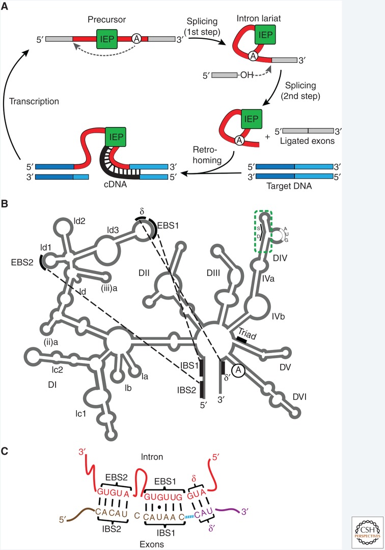
Figure 1.
Group II intron structure and function. (A) Intron life cycle. Splicing is initiated by attack on the 5′ splice site by the 2′-OH of the bulged adenosine (circled A) in DVI to form a lariat intermediate (first step). Attack on the 3′ splice site by the 3′-OH of the upstream exon results in ligated exons and the intron lariat (second step). The intron lariat can reverse splice into target DNA (retrohoming). The endonuclease of the intron-encoded protein (IEP) then nicks the opposite DNA strand, and the reverse transcriptase (RT) uses the cleaved DNA strand as a primer to make a cDNA copy of the integrated intron. After degradation of the intron RNA, second-strand DNA synthesis and repair, leading to a fully integrated DNA copy of the intron in the genome (not shown), transcription can ensue to complete the cycle. (B) Secondary structure of a group II intron. The six domains (DI–DVI) radiating from a central hub are labeled along with various subdomains, the “exon-binding sequence”–“intron-binding sequence” (EBS–IBS) pairings, the IEP anchor site in DIV (dashed green box), the catalytic triad in DV, and the bulged adenosine in DVI. SD, Shine–Dalgarno sequence. (C) EBS–IBS pairings. EBS1–IBS1, EBS2–IBS2, and δ-δ′ are shown. Blue dashed line denotes ligation site.

