Figure 2.
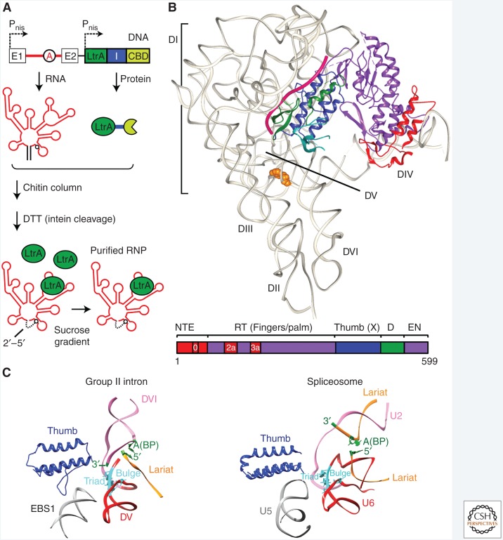
Cryo-electron microscopy (cryo-EM) structure of the Ll.LtrB group II intron. (A) Intein-based purification. The intron (red) and the intron-encoded protein (IEP) (termed LtrA, green) were expressed independently under nisin-inducible promoters (Pnis), with the IEP fused via an intein to a chitin-binding domain (CBD). The IEP promotes RNA splicing, and the resulting ribonucleoprotein (RNP) complex containing the IEP bound to excised intron lariat RNA was purified from lysed cells by binding to a chitin column. After dithiothreitol treatment, which induces intein cleavage, the released RNP was further purified on a sucrose gradient (Gupta et al. 2014; Qu et al. 2016). (B) Structure of the group II intron RNP determined by cryo-EM. The structure of the RNA (gray) and IEP (colored) was solved at a resolution of 3.8–4.5 Å. Cryo-EM was conducted by the laboratory of Dr. Hongwei Wang (Tsinghua University) and molecular interpretation of the cryo-EM map was a three-way collaboration among the Belfort and Wang laboratories and that of Dr. Raj Agrawal (Wadsworth Center) (Qu et al. 2016). RNA domains I–VI (DI–DVI) are shown (gray), along with the branch point adenosine (orange, space-filling) and trapped ligated exons (magenta). Below is a linear representation of the IEP, color-coded as in the cryo-EM structure, with amino-terminal extension (NTE), RT (fingers-palm), and thumb (X) domains, DNA-binding domain (D) and DNA endonuclease domain (EN). (C) Similarity of group II intron and spliceosome active centers. The group II intron (left) is shown alongside the Schizosaccharomyces pombe spliceosome (right) (Yan et al. 2015), illustrating the spatial similarity between the two. DV of the intron RNP and U6 of the U2/U6 spliceosome RNP (red) are aligned at their triad and bulge regions (cyan). The analogous structures of the group II intron and spliceosome are labeled: thumb domain in IEP and Prp8 (blue), the EBS1 loop of DI and U5 loop I (gray), DV and U6 of U2/U6 (red), DVI and U2 of U2/U6 (mauve), the lariat structures (orange), and their branch-point (BP) adenosines (green). (Adapted from Qu et al. 2016.)

