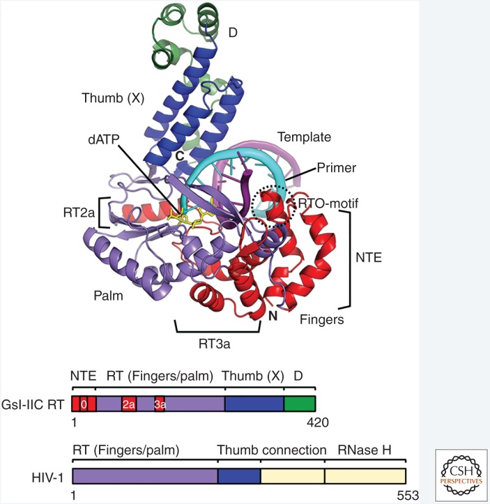
Figure 5.
Crystal structure of the thermostable GsI-IIC reverse transcriptase (RT) bound to template-primer and incoming dNTP. The 3.0 Å structure shows full-length GsI-IIC RT bound to an 11-bp RNA template-DNA primer duplex with a single-stranded 5′ overhang (3 nt) on the RNA template strand and a dideoxynucleotide at the 3′ end of the DNA primer. The incoming dATP is bound at the RT active site poised for polymerization. The amino-terminal extension (NTE) and insert regions RT2a and RT3a not present in retroviral RTs are demarcated with brackets. N and C indicate the amino- and carboxyl-termini of the protein. The schematics below show the domain organization of GsI-IIC RT compared with that of HIV-1 RT, with regions color-coded as shown in the figure. The connection and RNase H domains of HIV-1 RT not present in group II intron RTs are shown in yellow. (Adapted from Stamos et al. 2017.)

