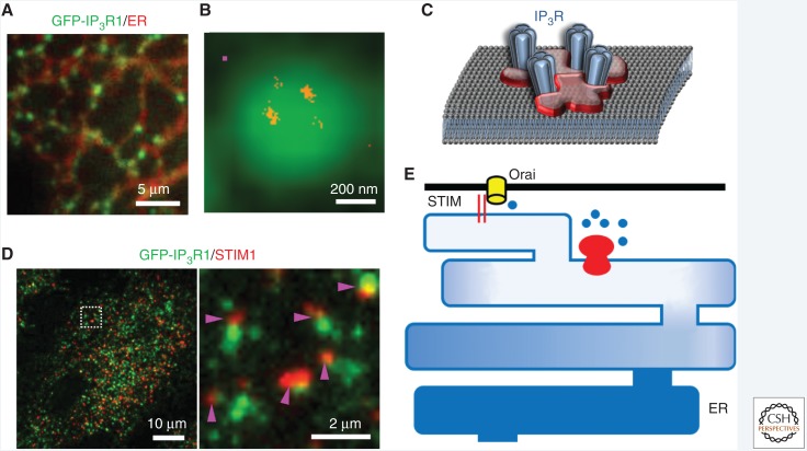
Figure 5.
Immobile IP3 receptor clusters initiate Ca2+signals. (A) HeLa cells with endogenous IP3R1 tagged with EGFP showing IP3R puncta (green) within endoplasmic reticulum (ER) membranes (red). (B) Diffraction-limited image of a punctum recorded using total internal reflection fluorescence (TIRF) microscopy, and the superimposed super-resolution (STORM) image showing IP3Rs (red) within the punctum. The magenta square indicates the approximate size of a single tetrameric IP3R. (C) IP3Rs appear to be diffusively distributed within a punctum, suggesting the need for a scaffold to corral IP3Rs. (D) TIRF images of a HeLa cell in which the ER has been depleted of Ca2+ by treatment with thapsigargin, showing the distribution of STIM1 (red) and IP3R (green). The enlarged image shows that stromal interaction molecule (STIM) puncta form alongside the ER where the immobile IP3Rs that are licensed to respond are parked. (Data results for A–D are from Thillaiappan et al. 2017.) (E) Licensed IP3Rs parked alongside the ER–PM junctions where store-operated Ca2+ entry (SOCE) occurs may allow substantial loss of Ca2+ from that part of the ER without causing appreciable Ca2+ loss from the remaining ER. The ER–PM junction with its associated licensed IP3Rs may constitute the basic functional unit for SOCE.

