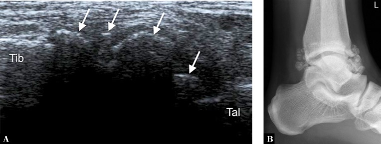
Fig. 4.
A. Longitudinal ultrasound through the anterior aspect of the ankle joint (Tib – anterior tibia, Tal – dorsal talus) of a 31-year-old man with synovial osteochondromatosis reveals multiple calcified loose bodies (arrows), corresponding to ossified osteochondral bodies seen on the lateral radiograph (B)

