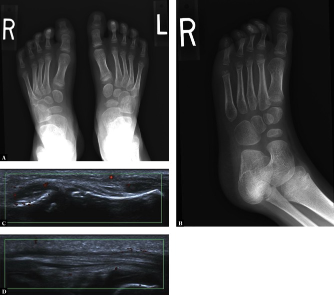
Fig. 6.
Right first toe synovitis and tenosynovitis in a 4-year-old girl: a feet AP radiograph (A) and an oblique view of the right foot (B) show increased density of periarticular soft tissues at the MTP1 and IP joints; ultrasound shows active synovitis at these 2 joints (C) and tenosynovitis of the FHL (D)

