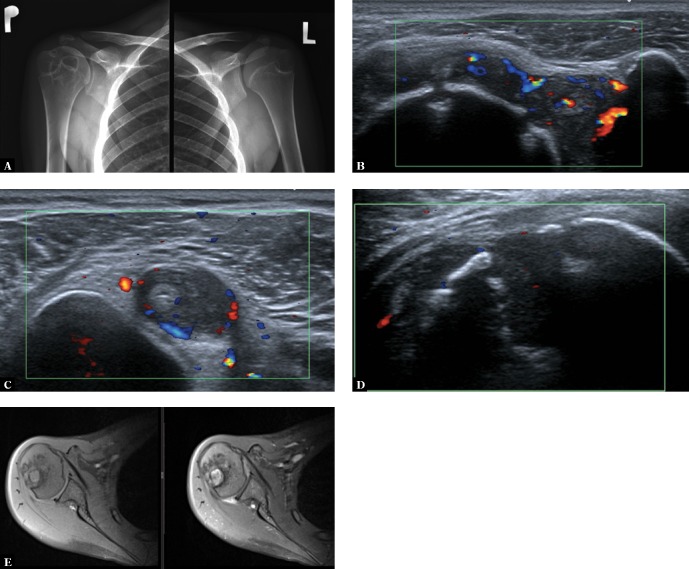
Fig. 7.
Glenohumeral joint US and MRI in a 17-year-old boy with Crohn’s Diseases: A. an AP radiograph shows numerous cysts in the right humeral head and medial aspect of the neck of the humeral bone, left joint is normal; ultrasound (B–D) shows active synovitis and two small erosions of the humeral head (B), tenosynovitis of the long head of the biceps tendon and (C), deep inactive erosion of the humeral head (D); an MRI T1-weighted fat-suppressed (FS) (E) and postcontrast T1-weighted FS (F) images show BME, a large cyst, multiple erosions, and a postcontrast signal increase within inflammatory lesions

