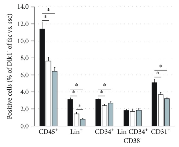
Figure 8.

Flow cytometry analysis after Dlk1+ cell depletion. Flow cytometry analysis of Dlk1-negative human fetal liver-derived cells after five days in culture with Dlk1+ cells in direct contact (black bars), without Dlk1+ cells (white bars), or with Dlk1+ cells in inserts (grey bars). Data are given as means from n = 4 biological repeats ± standard deviation. ∗ indicates a statistically significant difference (p ≤ 0.05). Abbreviations: fsc: forward scatter; ssc: side scatter; CD: cluster of differentiation.
