Fig. 3.
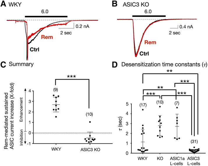
Effect of Rem on sustained ASIC currents in acutely isolated Dil-labeled DRG neurons from WKY (control) and ASIC3 KO rats. (A and B) Representative ASIC current traces obtained from DRG neurons from WKY (A) and ASIC3 KO (B) rats before (Ctrl [control], black) and after Rem (10 μM, red) exposure. The solid bars above the traces represent a 10-second exposure to the pH 6.0 test solution. The neurons were preexposed to Rem for 3 minutes (pH 7.4) before exposure to the test solutions (pH 6.0 + Rem). (C) Summary dot plot indicating the mean (± S.D.) Rem-mediated sustained ASIC current enhancement (X-fold). ***P < 0.0001 using unpaired two-tailed t test. (D) Mean (± S.D.) desensitization time constant (τ) values measured for ASIC currents in DRG neurons isolated from WKY rats (control), ASIC3 KO rats, and L-cells transfected with either ASIC1a or ASIC3 cDNA. The holding potential (VH) for DRG neurons and L-cells was −80 and −60 mV, respectively. Numbers in parenthesis indicate the number of recordings. ***P < 0.0001 and **P < 0.001, respectively, using one-way ANOVA followed by Tukey post hoc test.

