Figure 8.

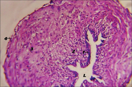
Photomicrograph showing the histological structure of the 1-week-old chick left oviduct. Inner epithelial layer (1), mesenchymal cell layer (2), circular smooth muscle layer (3), tunica serosa (4), folded lumen (H and E, ×40). L: Lumen

Photomicrograph showing the histological structure of the 1-week-old chick left oviduct. Inner epithelial layer (1), mesenchymal cell layer (2), circular smooth muscle layer (3), tunica serosa (4), folded lumen (H and E, ×40). L: Lumen