Figure 9.

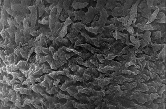
Scanning electron micrograph showing the tunica mucosa of the 1-week-old chick left oviduct with extensive mucosal folds and many corrugated cilia (×5000)

Scanning electron micrograph showing the tunica mucosa of the 1-week-old chick left oviduct with extensive mucosal folds and many corrugated cilia (×5000)