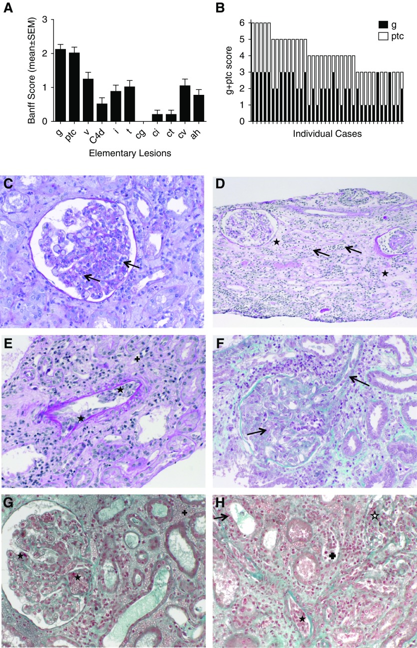
Figure 2.
Representative pathologic characteristics of the early AMVRs. (A) Mean (±SEM) values of the elementary lesions assessed using the Banff classification in the biopsy samples from 38 KTRs at time of AMVR. (B) The glomerulitis and peritubular capillaritis (g+ptc) scores of the 38 individual patients with AMVR. (C) Image of periodic acid–Schiff (PAS) staining showing severe glomerulitis with partial to complete occlusion of glomerular capillaries by infiltrating leukocytes (mononuclear cells and neutrophils cells) (arrows). (D) Image of PAS staining showing severe peritubular capillaritis (ptc3) with more than ten inflammatory cells in dilated capillaries (arrows) associated with diffuse interstitial edema (+). (E) Image of PAS staining showing intimal arteritis v2 with mononuclear cells underneath the endothelium and occlusion of more than 25% of the arterial lumen (⋆) associated with peritubular capillaritis (+) and sparse inflammatory cells within the interstitium. (F) Image of Masson trichrome staining showing severe glomerulitis with complete occlusion of glomerular capillaries by infiltrating leukocytes and EC enlargement. EC enlargement is also present in arterioles (arrows). (G) Image of Masson trichrome staining showing thrombotic microangiopathy characterized by thrombi in the glomerular capillaries (⋆) associated with glomerulitis, peritubular capillaritis, and diffuse interstitial hemorrhage (+). (H) Image of Masson trichrome staining showing a mixed rejection with diffuse interstitial inflammation and tubulitis (arrow), glomerulitis open star, peritubular capillaritis (+), arteriolitis (⋆), and interstitial hemorrhage.

