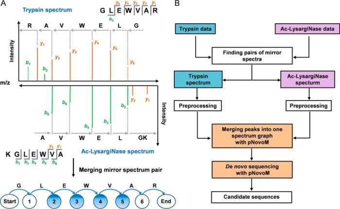
Fig. 2.
Mirror protease strategy for de novo sequencing. A, The mirror spectra improved the quality of the matched peptide-spectrum pairs by providing a continuous and complete set of product ions and distinguishable directions of most ions (marked with blue circles, e.g. vertices 2, 3, 4 and 5). Two dotted peaks denote that b1 and y6 were missing in the trypsin spectrum. B, Workflow of the pNovoM algorithm based on the mirror protease strategy for de novo sequencing. Mirror spectrum pairs from trypsin- and Ac-LysargiNase-digested samples were found and sequenced using pNovoM. Then, the two spectra in each pair were integrated into one merged spectrum graph for de novo sequencing.

