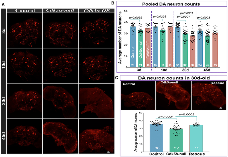
Figure 1. Both Gain and Loss of Cdk5α Induce the Degeneration of Dopaminergic Neurons.
Brains of 3-, 10-, 30-, and 45-day-old flies of the indicated genotypes were dissected and immunostained with anti-DTH antibody.
(A) Representative projected confocal images of DA neurons labeled with anti-DTH antibody.
(B) The number of DTH+ DA neurons per hemisphere is presented as mean ± SEM, along with individual counts. The pooled DA neuron count includes neurons from PPL1, PPL2, PPM1/2, PPM3, and PAL clusters. For individual counts in these clusters, see Figure S1. For each genotype and age, the number of hemispheres examined is presented at the bottom of the bar. Error bars indicate SEM. The significance of differences is relative to the age-matched control (one-way ANOVA with Dunnett’s multiple correction). The DA neuron count in 45-day-old Cdk5α-OE shows an apparent rebound in numbers relative to 30 days; this reflects selective survival of only the fittest individuals at the oldest time point (Spurrier et al., 2018). For complete details of statistical analysis for this and all of the figures, see Table S2.
(C) Top: projected confocal images of 30-day-old brains of DTH-stained control, Cdk5α null, and Rescue {Cdk5α null; Tn[Cdk5α]/+}. Bottom: counts showing mean ± SEM along with individual values. For the rescue samples, the significance of differences was calculated between rescue and age-matched Cdk5α null using one-way ANOVA with Tukey’s multiple correction. In the rescue calculation, the same data for 30-day-old controls and Cdk5α null were used.

