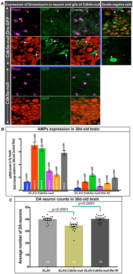
Figure 3. Cdk5α-Altered Drosophila Have Neuronal Expression of AMPs.
(A) Projected confocal image showing drosomycin expression (green) in the brains of 30-day-old Cdk5α null Drosophila along with embryonic lethal, abnormal vision (ELAV) (red, neuronal marker) and reversed polarity (Repo) (magenta, glial marker). Yellow arrowheads highlight drosomycin in glial cells, while white arrows highlight expression in neurons. Some GFP+ cells were observed that were positive for neither neuronal nor glial markers (stars); some of these resemble hemocytes. Five biological replicates were examined.
(B) AMP expression in Cdk5α null flies with or without Elav-GAL4-mediated knock down of Relish expression. Fold change of AMPs was calculated versus 30-day-old controls and presented as mean ± SEM; significance in Elav-GAL4; Cdk5α null was assessed for each AMP by comparison to age-matched Elav-Gal4, while for Elav-Gal4;Cdk5α null; Relish RI, it was compared to Elav-Gal4;Cdk5α null, using Student’s t test for five biological replicates.
(C) DA neuron counts in 30-day-old Elav-Gal4, Elav-Gal4;Cdk5α null, and Elav-Gal4;Cdk5α null;Relish RI. Flies were aged at 25°C, and DA neurons were counted by staining with anti-TH antibody. Data are presented as mean ± SEM with individual counts shown. The number of brain hemispheres counted are at the bottom of each bar. Statistical significance was determined using one-way ANOVA with Tukey’s multiple correction.

