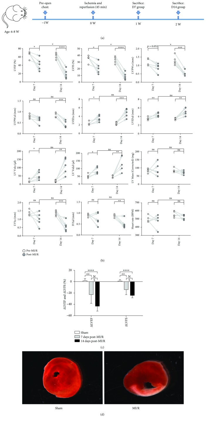
Figure 1.
Cardiac dysfunction in mice post-MI/R. (a) The main time shaft of animal experiments was presented. (b) LVFS (%), LVPW (mm), LVID (mm), LV Vol (μl), LV Mass (corrected) (mg), IVS (mm), and heart rates (BPM) were measured under transthoracic echocardiography pre-MI/R and post-MI/R at day 7 and day 14, respectively. ∗P < 0.05, ∗∗P < 0.01, ∗∗∗P < 0.001, and ∗∗∗∗P < 0.0001. (c) ΔLVEF (difference mean value between pre-MI/R and post-MI/R) and ΔLVFS (difference mean value between pre-MI/R and post-MI/R) were measured among the three groups. ∗∗P < 0.01 and ∗∗∗∗P < 0.0001. (d) The heart tissues were stained by TTC.

