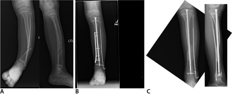
Fig. 5.
Anteroposterior (AP) (left) and lateral (right) radiographs (a) of left tibia and fibula with anterolateral bowing, neurofibromatosis and tibial pseudarthrosis with an intact fibula. This is classified as Paley type 3. AP radiograph (b) six months after cross-union surgery performed at age five years. AP radiograph (c) one and a half years after cross-union surgery. The plate has been removed. The rod was exchanged for a new Fassier-Duval rod.

