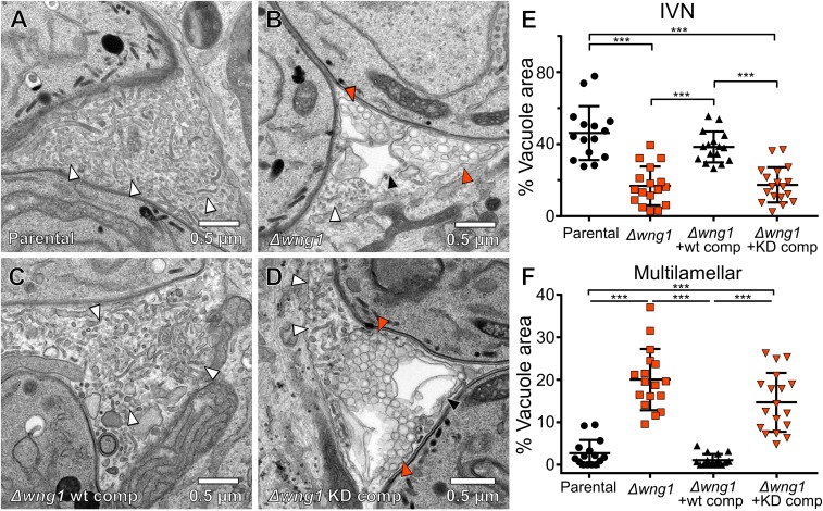
Fig. 5.
Vacuoles lacking active WNG1 kinase show disrupted IVN membranes. Representative transmission electron microscopic images of the (A) parental, (B) RHΔwng1, (C) RHΔwng1 complemented with WT WNG1, and (D) kinase-dead complemented strains. IVN tubules are indicated with white arrowheads. Multilamellar vesicles are indicated with solid orange arrowheads. Multilamellar structures in which internal vesicles appear to have been lost during fixation are indicated with a black arrowhead in B and D. The relative area of each IVN tubules and multilamellar vacuole from EM images as in A–D were quantified in ImageJ. (E and F) Significance was calculated in Prism by ANOVA with Tukey’s test; ***P < 0.0001. Note that the parental vs. WT-comp and Δwng1 vs. KD comp showed no significant difference.

