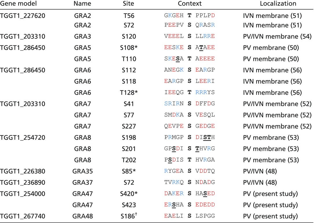
Table 1.
Phosphosites down-regulated in RHΔwng1 vacuoles
 |
The sequence context of each of the phosphosites is indicated. Acidic residues are red, basic residues are blue. Note that some regions appear to be hyperphosphorylated in a WNG1-dependent manner. Such potential priming sites are indicated bolded, with bolded and underlined in the phosphosite context.
*Not significant in t test due to variability between replicates (P > 0.05) or quantified in only one type of labeling.
†Identified in preliminary dataset, not statistically significant in final data.
