Fig. 1.
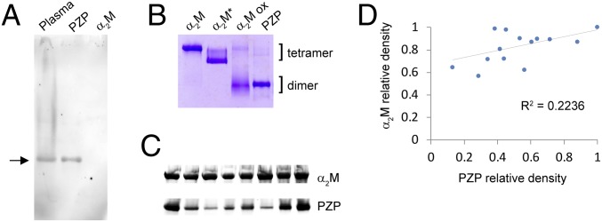
Western blot and native PAGE analyses of PZP in human pregnancy plasma and following purification. (A) Image of a Western blot showing the migration of PZP pre- and postpurification from pregnancy plasma, after separation using a 3 to 8% Tris-acetate native gel. The position of PZP is indicated with an arrow. A corresponding amount of purified α2M was not detected (final lane). (B) Image of a 3 to 8% Tris-acetate native gel showing the migration of purified PZP, α2M, transformed α2M (an electrophoretically fast tetramer), and hypochlorite-modified α2M (α2M ox). The positions of αM tetramers and dimers are indicated. (C) Images of Western blots showing the relative PZP levels in eight individual pregnant women at 36 wk gestation. Pregnancy plasma (1 µL) was separated using a 3 to 8% Tris-acetate native gel, and proteins were transferred to a nitrocellulose membrane. The blot was initially probed using an anti-PZP antibody, and then the same blot was reprobed using an anti-α2M antibody. Images show the major bands detected, and are cropped and realigned to assist comparison of the respective levels of PZP and α2M present. (D) Densitometry analysis of Western blots showing the relative levels of PZP and α2M in 14 individual women at 36 wk of pregnancy. R2 is calculated using Pearson’s correlation coefficient.

