Fig. 5.
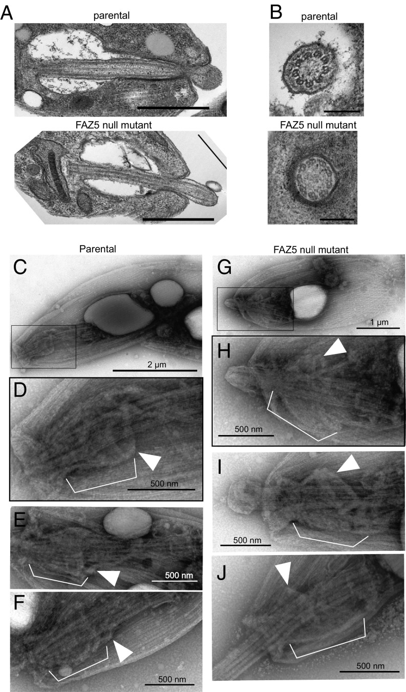
Deletion of FAZ5 dramatically alters the FP architecture in amastigotes. (A) Electron micrographs of longitudinal sections through the FP of parental and FAZ5 null mutant axenic amastigotes. (Scale bars, 500 nm.) (B) Electron micrographs of cross-sections through the axoneme, showing the lack of central pair microtubules. (Scale bars, 200 nm.) Whole-mount cytoskeletons of parental (C–F) and FAZ5 null mutant (G–J) axenic amastigotes were subjected to negative staining for observation by TEM. (C and G) Images of whole cells (with Insets in D and H, respectively) showing the position of the cytoskeletal structures displayed in higher magnification images. At the neck region of amastigote cytoskeletons, the axoneme is surrounded by a complex structure (white brackets). At its proximal end (relative to the flagellum base), this structure is delimited by filaments likely to correspond to the FP collar (white arrowheads). In parental cells, the neck cytoskeleton has a wine glass shape. In contrast, the neck cytoskeleton of FAZ5 null mutants appeared cup-shaped, with a considerably wider FP collar than that of parental cytoskeletons.

