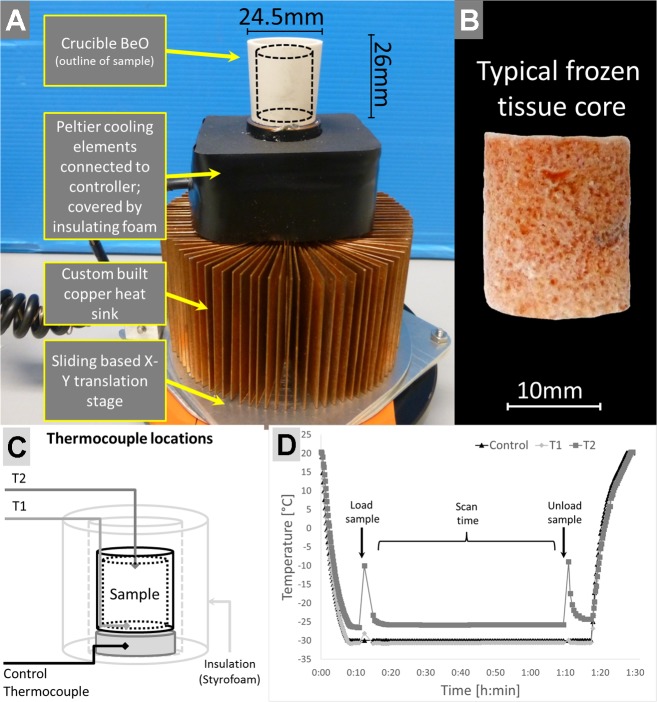
Fig. 2.
Cooling stage design, example sample, and testing. A: the cooling stage is composed of a beryllia crucible mounted on a small copper stub which enables the Peltier elements to draw heat away from the sample. The Peltier elements are directly mounted on top of a custom-made heat sink. The entire assembly is mounted on an X-Y translation stage which enabled a precise positioning in the scanner. B: a typical frozen lung tissue core which was extracted from a whole air-inflated frozen lung; the diameter is ~16 mm, and it is 20-mm high. C: schematic of stage indicating the location of additional temperature probes (T1, T2) within the sample holder. D: temperature plot over the duration of one experiment including precooling time, scan time, and warm-up phase after scan completion and sample removal.

