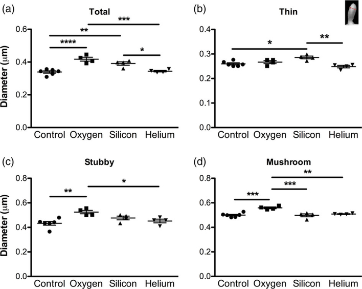
FIGURE 4.
Charged particle irradiation results in changes in spine morphology. Analysis of spine head diameter in CA1 apical dendritic spines. (a) Average total spine head diameter, (b) thin spine head diameter, (c) stubby spine head diameter and (d) mushroom spine head diameter. Inset image in b shows how spine head diameter was assessed. Data represent means ± SEM. One-way ANOVA *p < 0.05, **p < 0.01, ***p < 0.001, ****p < 0.0001 [Color figure can be viewed at wileyonlinelibrary.com]

