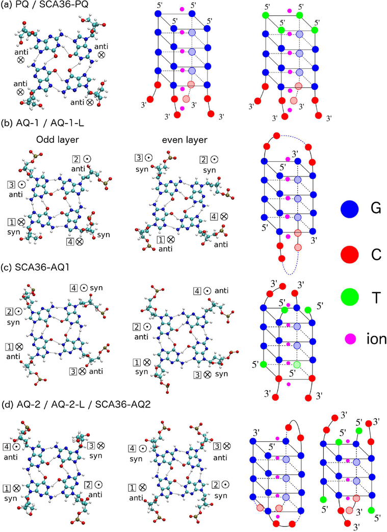
Figure 1.
Quadruplex geometries. Left: Geometries of guanine residues within a quartet (odd layers in first column and even layers in second column). Right: full quadruplex configurations. (a) PQ and SCA36-PQ; (b) AQ-1/AQ-1-L; (c) SCA36-AQ1 (d) AQ-2/AQ-2-L/SCA36-AQ2. G’s have been labeled as blue, C’s are red, T’s are green, and ions are magenta. ⊗ indicates 5′ → 3′ strand direction pointing into the paper; ⊙indicates 5′ → 3′ strand direction pointing out of the paper.

