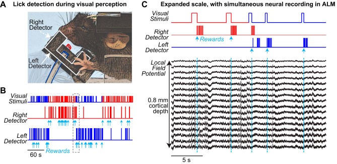
Fig. 8.
Dual-port detection during behavior with simultaneous neural recording. A: fully assembled detector during behavioral sessions in a head-fixed mouse. White outline indicates left detector, colored outlines indicate silicone tubing for water delivery at left (blue) and right (red) detector ports. Right detector is invisible in this view. B: example behavioral session, from a different mouse. Top row shows onsets of visual stimuli in left (blue) or right (red) visual fields. Bottom 2 traces show licks detected at each sensor. Individual licks are not visible at this scale. The mouse reports perception of stimulus onset in each location by licking at the appropriate detector. Cyan arrows indicate reward delivery at each individual detector (one reward per correct trial). C: expanded view of session in B, with simultaneous neural recording from anterolateral motor cortex (ALM). Mouse performs 2 correct trials of rightward detection, and then switches to leftward detection on third trial after a few erroneous licks. Local field potential (LFP) recorded with a multisite linear silicon probe throughout the cortical depth of ALM (0.8 mm below surface, 50-μm spacing between contacts) shows no artifacts during licking (see Fig. 9).

