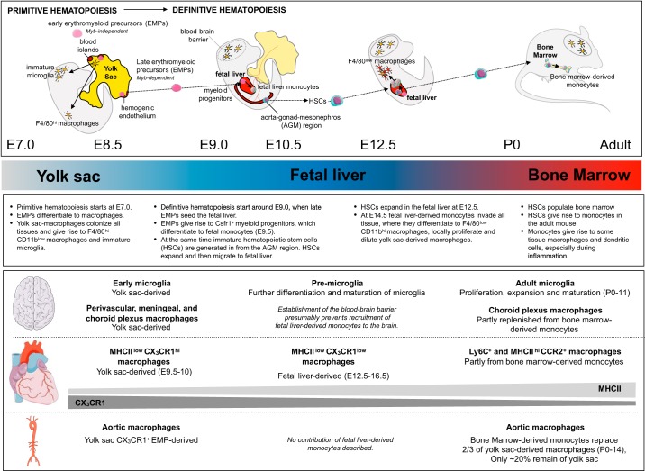
FIGURE 2.
Macrophage origin in the brain and cardiovascular organs. The cartoon depicts important steps in the development of monocytes and tissue-resident macrophages. The main organ of hematopoiesis (yolk sac, fetal liver, and bone marrow) is indicated. The main steps in tissue-resident macrophage ontogeny, as well as the origin of specific macrophage subsets in brain, heart, and aorta, are highlighted below the schematic. Density gradients of major histocompatibility complex II (MHCII) and CX3CR1 in cardiac macrophages indicate that these decrease or increase with age, respectively. Overall proliferation capacity of cardiac macrophages decreases with age, and they are increasingly replenished by monocytes. AGM, aorta-gonad-mesonephros; E, embryonic day; EMPs, early erythromyeloid precursors; HSCs, hematopoietic stem cells. P, postnatal day.

