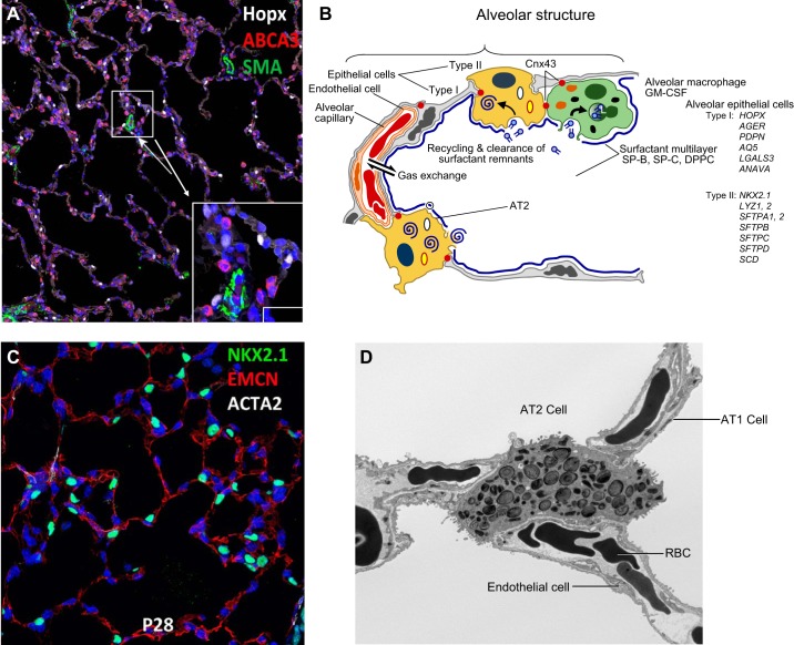
FIGURE 11.
Structure of the alveolar gas-exchange region. A: gas exchange is facilitated by creation of a vast surface area lined primarily by AT1 cells (HOPX, in white) and AT2 cells (ABCA3 in red) or NKX2–1 in green (C). AT2 cells synthesize and secrete pulmonary surfactant lipids and proteins that reduce surface tension in the alveoli after birth. SMA (green) marks vascular smooth muscle. B: surfactant components are secreted into the alveoli and recycled by AT2 epithelial cells. Surfactant is catabolized by alveolar macrophages by processes regulated by GM-CSF. Proteins selectively expressed by AT1 or AT2 cells are listed on the right of the panel. D: an electron micrograph demonstrates lamellar bodies containing surfactant lipids and proteins in AT2 epithelial cells. AT1 cells line the majority of the alveolar gas exchange surface, coming into close contact with endothelial cells in the pulmonary microvasculature to facilitate transport of O2 and CO2 to red blood cells (RBC). [Modified from Whitsett and Alenghat (353).]

