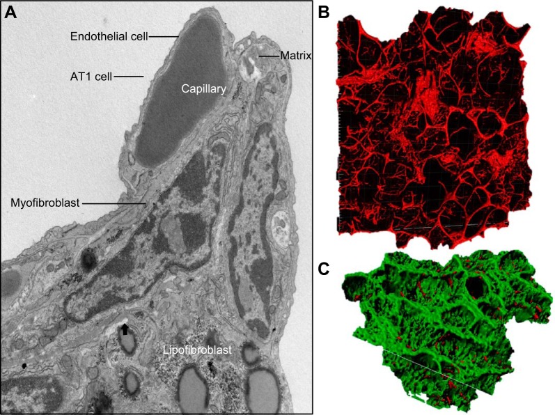
FIGURE 17.
Diverse fibroblasts build the alveolar scaffold. A: the electron micrograph shows the ultrastructure of an alveolar septae from the adult mouse lung. Most of the alveolar surface is covered by AT1 epithelial cell(s). Gas exchange occurs across the AT1-endothelial interface with alveolar capillaries. Matrix-, lipo-, and myofibroblasts are seen in the septal wall. A collagen-elastin bundle (matrix) is seen at the septal tip. B and C: confocal imaging of the mouse lung (PND3) is shown after injection with isolectin B4 visualizing endothelial cells of the alveolar capillary network. Surfaces of the alveoli were rendered from confocal images after staining the endothelium with isolectin B4 lectin after acquisition at 585–635 nm (green). Second harmonic imaging shows collagen bundles in red. (Figure courtesy of Dr. Matt Kofron, used with permission).

