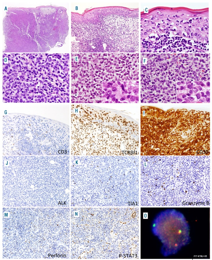
Figure 3.
Histological and immunophenotypic features of a primary cutaneous anaplastic large cell lymphoma with DUSP22 rearrangement (case 5). (A) Low-power microscopic image of the skin biopsy showing diffuse dermal infiltration, characterized histologically by a dense dermal infiltrate with epidermal involvement by small lymphocytes (B and C). (D) Dermal infiltrate of medium-sized and atypical lymphocytes, with a monomorphic appearance, including hallmark and occasional doughnut cells (E, inset; F, inset). Neoplastic cells were CD3-negative (G), TCRβF1-positive (H), and CD30-positive (I). ALK (J), TIA-1 (K), granzyme B (L), and perforin (M), and P-STAT3 (N) were negative. (O) Fluorescence in situ hybridization using a break-apart probe at the DUSP22 locus shows a rearrangement, with one normal fusion signal and an abnormal split signal.

