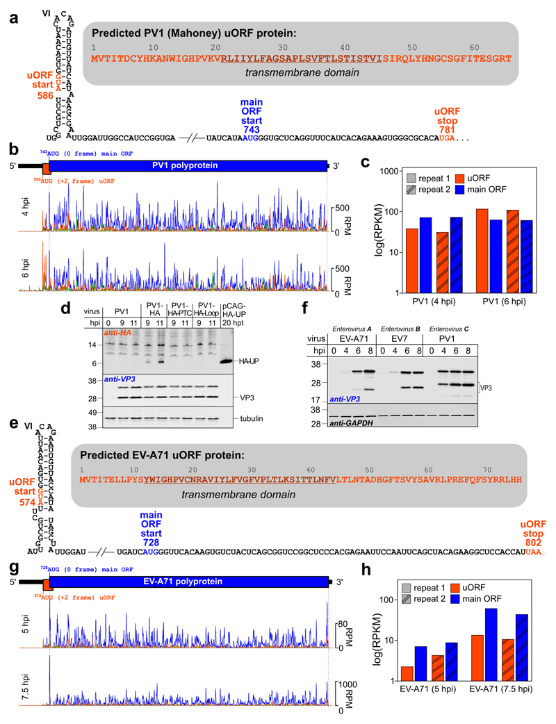
Fig. 4. Translation of the uORF in poliovirus PV1 and enterovirus EV-A71.
a, Schematic representation of the PV1 IRES dVI and uORF region with the uORF start and stop (orange) and main ORF start (blue) annotated. The UP amino acid sequence is shown in the shadowed inset with the predicted TM domain underlined. b, Ribosome profiling of PV1-infected cells at 4 and 6 hpi. Ribo-Seq RPF densities in reads per million mapped reads (RPM) are shown with colors indicating the three phases relative to the main ORF (blue – phase 0, green – phase +1, orange – phase +2), each smoothed with a 3-codon sliding window (see Fig. S12a for repeats). c, Mean ribosome density in the PV1 uORF and main ORF at 4 and 6 hpi, based on the in-phase Ribo-Seq density in each ORF (excluding the overlapping region; RPKM = reads per kilobase per million mapped reads). d, Analysis of viral protein expression in RD cells infected with wt or HA-tagged PV1 viruses. Cells were infected at an MOI of 50, harvested at 9 and 11 hpi, and accumulation of HA-tagged UP (HA-UP) and virus structural protein VP3 was analyzed by western blotting with anti-HA, anti-VP3 and anti-tubulin antibodies. HA-UP transiently expressed from a pCAG promoter in HeLa cells taken at 20 h post-transfection was used as a HA-UP size control. e, Schematic representation of the EV-A71 IRES dVI and uORF region with the uORF start and stop (orange) and main ORF start (blue) annotated. The UP amino acid sequence is shown in the shadowed inset with the predicted TM domain underlined. f, Analysis of protein expression in RD cells infected with enteroviruses EV-A71, EV7 or PV1. Cells were infected at an MOI of 20, harvested at 0–8 hpi as indicated, and expression of virus and host proteins was analyzed by western blotting with anti-VP3 and GAPDH antibodies. The experiments in (d,f) were independently repeated three times with similar results. g, Ribosome profiling of EV-A71-infected cells at 5 and 7.5 hpi (see Fig. S12b for repeats). h, Mean ribosome density in the EV-A71 uORF and main ORF at 5 and 7.5 hpi.

