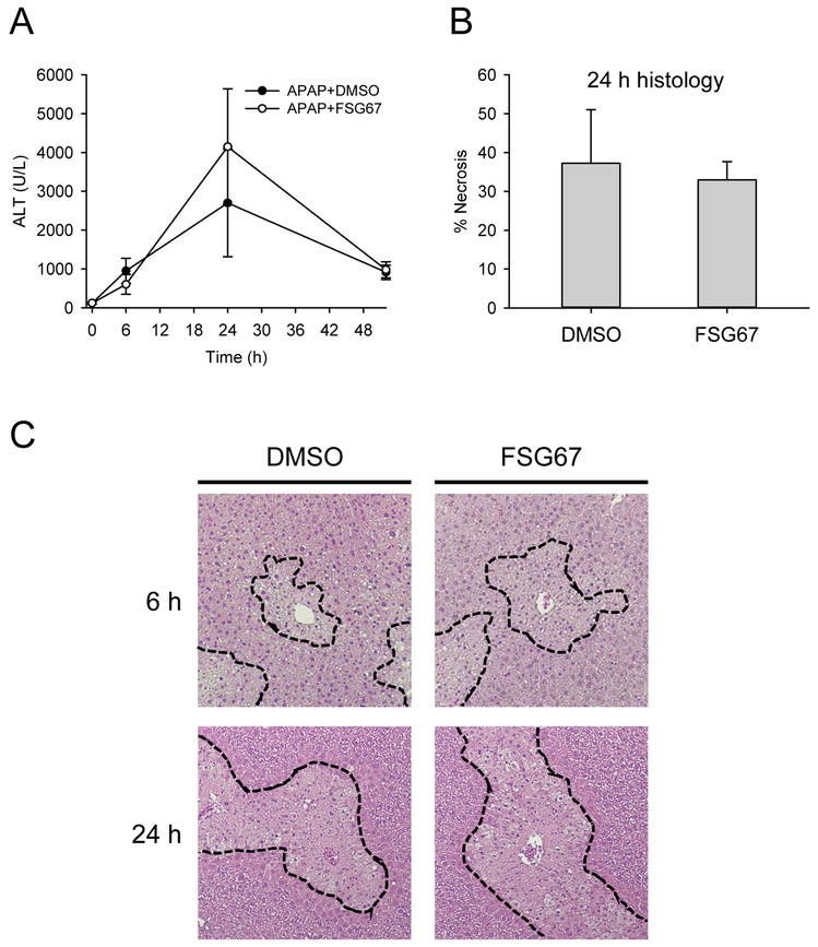
Figure 1. FSG67 does not affect liver injury after APAP overdose.
Mice were treated with 300 mg/kg APAP at 0 h followed by either DMSO vehicle or 20 mg/kg FSG67 at 2, 24, and 48 h. Blood and liver tissue were collected at 6, 24 and 52 h. (A) Plasma alanine aminotransferase (ALT) activity over time. (B) Percentage of tissue that was necrotic at 24 h. (C) H&E-stained liver sections at 6 and 24 h. Dashed lines emphasize areas of necrosis. Necrosis is characterized by loss of basophilia, increased eosinophilia, and nuclear shrinkage (pyknosis) at 6 h, and by eosinophilia, cell swelling, and loss of nuclei (karryorhexis) at 24 h. Data expressed as mean±SE for n = 3-6 mice.

