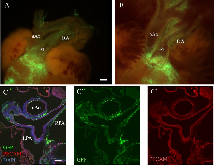
Fig 3. Distribution of Tbx1-expressing cells and their descendants in mouse fetuses.
A.B: Whole mount fluorescent photographs of the outflow region of a E18.5 Tbx1cre/+; RosamT/mG fetus. A: external appearance. B: internal optical plane. Note the heavy contribution of GFP+ cells to the pulmonary myocardium, pulmonary trunk (PT) and pulmonary valves, but superficial contribution (endothelial and adventitial) to other great vessels, including the ductus arteriosus (DA). which appears to have a more dense endothelial contribution. aAo: ascending aorta. C: immunofluorescence of a transverse section of a E18.5 Tbx1cre/+; RosamT/mG. Anti GFP staining is shown in green, anti PECAM1 staining (endothelial-specific) is shown in red. DAPI staining (cell nuclei) is shown in blu. C'-C'': green and red channels are shown separately. aAo: ascending aorta; PT: pulmonary trunk and pulmonary leaflets; LPA, RPA: left and right pulmonary arteries. Scale bar is 50 micrometers in A and B, 100 micrometers in C-C".

