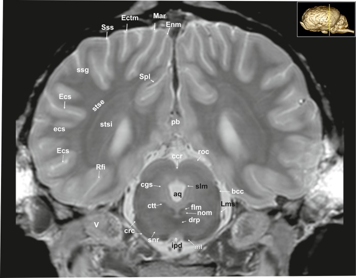
Fig 10. Transverse magnetic resonance image of the equine brain on the level of the rostral colliculi.
aq: mesencephalic aqueduct, bcc: brachium of the caudal colliculus, ccr: commissure of the rostral colliculus, cgs: central grey substance, crc: cerebral crus, ctt: central tegmental tract, drp: decussation of the rostral cerebellar peduncles, Ecs: ectosylvian sulcus, Ectm: ectomarginal sulcus, Enm: endomarginal sulcus, flm: medial longitudinal fasciculus, ipd: interpeduncular nucleus, Lms: lateral mesencephalic sulcus, Mar: marginal sulcus, ml: medial lemniscus, nom: nucleus of the oculomotor nerve, pb: pineal body, Rfi: rhinal fissure, roc: rostral colliculus, snr: substantia nigra, Spl: splenial sulcus, stse: stratum sagittale externum, stsi: stratum sagittale internum, Sss: suprasylvian sulcus, V: trigeminal nerve.

