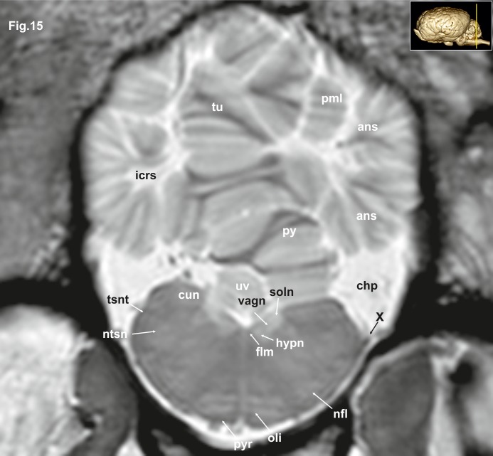
Fig 15. Transverse magnetic resonance image of the equine brain on the level of the cuneate nuclei.
ans: ansiform lobule, chp: choroid plexus; cun: cuneate nucleus, flm: medial longitudinal fasciculus, hypn: nucleus of the hypoglossal nerve, icrs: intercrural sulcus, nfl: nucleus of the lateral fascicle, ntsn: nucleus of the spinal tract of the trigeminal nerve, oli: olivary nucleus, pml: paramedian lobule, py: pyramis of the vermis, pyr: pyramidal tract, tsnt: spinal tract of the trigeminal nerve, tu: tuber of the vermis, uv: uvula of the vermis, vagn: nucleus of the vagus nerve, X: vagus nerve.

