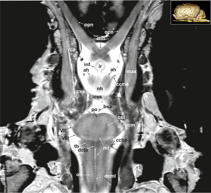
Fig 17. Dorsal magnetic resonance image of the equine brain on the level of the optic chiasm.
ah: adenohypophysis, bsa: basilar artery, ccba: caudal cerebellar artery, ccma: caudal communicating artery, cho: optic chiasm, cst: corticospinal tract, cvs: cavernus sinus, dcml: decussation of medial lemniscus, dctb: decussation of the fibres of trapezoid body, icvs: intercavernous sinus, inf: infundibular stalk, ir: infundibular recess, max: maxillary nerve, ml: medial lemniscus, nh: neurohypophysis, oli: olivary nucleus, opn: optic nerve, pcm: peduncles of the mammillary body, po: pons, rca: rostral cerebral artery, tb: trapezoid body, VII: facial nerve, VIII: vestibulocochleal nerve.

