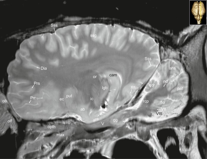
Fig 32. Parasagittal magnetic resonance image of the equine brain at the level of the optic radiation.
ab: amygdaloid body, ans: ansiform lobule, are: entorhinal aera, cam: ammon’s horn, chp: choroid plexus, Dia: diagonal sulcus, ec: external capsule, flo: flocculus, gg: Gasserian ganglion, ic: internal capsule, lgb: lateral geniculate body, max: maxillary nerve, mgb: medial geniculate body, nd: dentate nucleus, ob: olfactory bulb, olf: olfactory fibres, opth: ophthalmic nerve, or: optic radiation, paf: paraflocculus, Prr: prorean sulcus, Prs: presylvian sulcus, put: putamen, qdr: quadrangulare lobule, rcp: rostral cerebellar peduncle, Rfi: rhinal fissure, ssg: suprasylvian gyrus, Sss: suprasylvian sulcus, V: trigeminal nerve, VII: facial nerve, VIII: vestibulocochleal nerve.

