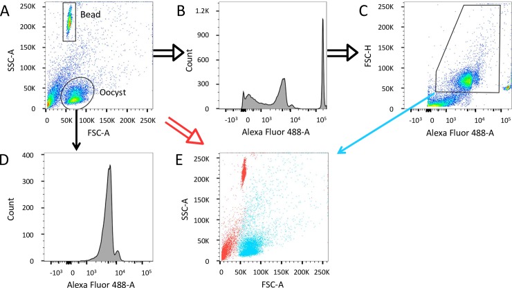
Fig 1. Staining of oocysts with a mouse anti-Cryptosporidium Alexa Fluor 488 monoclonal antibody.
(A to E) Representation of an Alexa 488 stained sample containing 104 oocysts. (A) A representative plot of oocysts purified from mouse intestine. The oval Oocyst gate encompasses the expected location of the C. parvum oocysts on the plot of SSC-A versus FSC-A based on a positive control of purified oocysts. Histograms showing the distribution of events positive for Alexa Fluor 488 emission on the ungated sample (B) and the Oocyst gate (D). (C) A pseudo-coloured dot plot of FSC-H versus Alexa 488 with the recommended gate for Alexa 488+ oocysts. (E) Overlay of Alexa 488+ oocysts from panel C (blue) and negative events (red) of the ungated sample.

