-
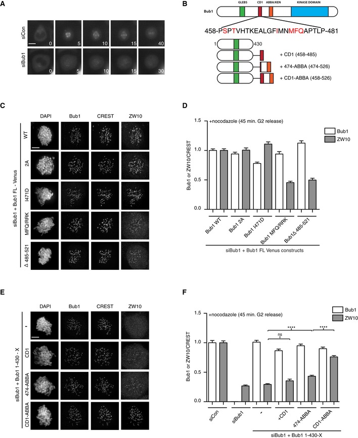
A
Localization of Venus‐Rod during unperturbed mitosis in control‐depleted or Bub1‐depleted cells.
-
B
Primary structure of Bub1 with sequence of CD1 shown and residues mutated in red. Schematic of fusion constructs used in panels (E and F).
-
C
HeLa cells were depleted of Bub1 using RNAi and complemented with indicated Venus‐Bub1 constructs. Immunofluorescence images were shown for Bub1 and ZW10 localization.
-
D
Quantification of Bub1 and ZW10 kinetochore levels normalized to CREST in the indicated conditions.
-
E, F
As in (C and D) but with the indicated Bub1 fusion proteins (Student's t‐test used for statistical comparison, ns: non‐significant, ****P ≤ 0.0001).
Data information: Bar indicates mean and standard error of mean is shown by line. At least 200 kinetochores from 10 cells were analyzed and representative result from at least two independent experiments is shown. Scale bars, 5 μm.