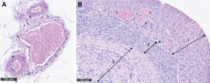
Figure 1.
(A) Cross sections of the uterus showing the veins (v) and arteries (a) within the mesometrium stained with H&E. Corresponding to the two uterine horns there are two main utero-ovarian arteries and veins that are positioned in the mesometrium parallel to each uterine horn. Arcuate vessels run from the main vessels and form loops that are also positioned in the mesometrium. (B) At the same magnification, a cross section through the uterus provides a view on a fraction of the uterus showing the myometrial vessels (radial, a, v) connecting the arcuate vessels to the uterine wall. Arrow 1 marks the myometrium, and arrow 2 indicates the endometrium.

