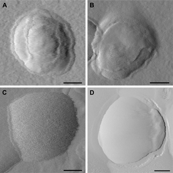
Figure 3.
Representative AFM images of OTR-Lipo (A), ATO-Lipo (B), rabbit IgG immunoliposomes (C), and conventional liposomes (D).
Note: Scale bars correspond to 50 nm.
Abbreviations: AFM, atomic force microscopy; OTR-Lipo, PEGylated immunoliposomes conjugated with anti-oxytocin receptor monoclonal antibodies; ATO-Lipo, atosiban-conjugated PEGylated liposomes.

