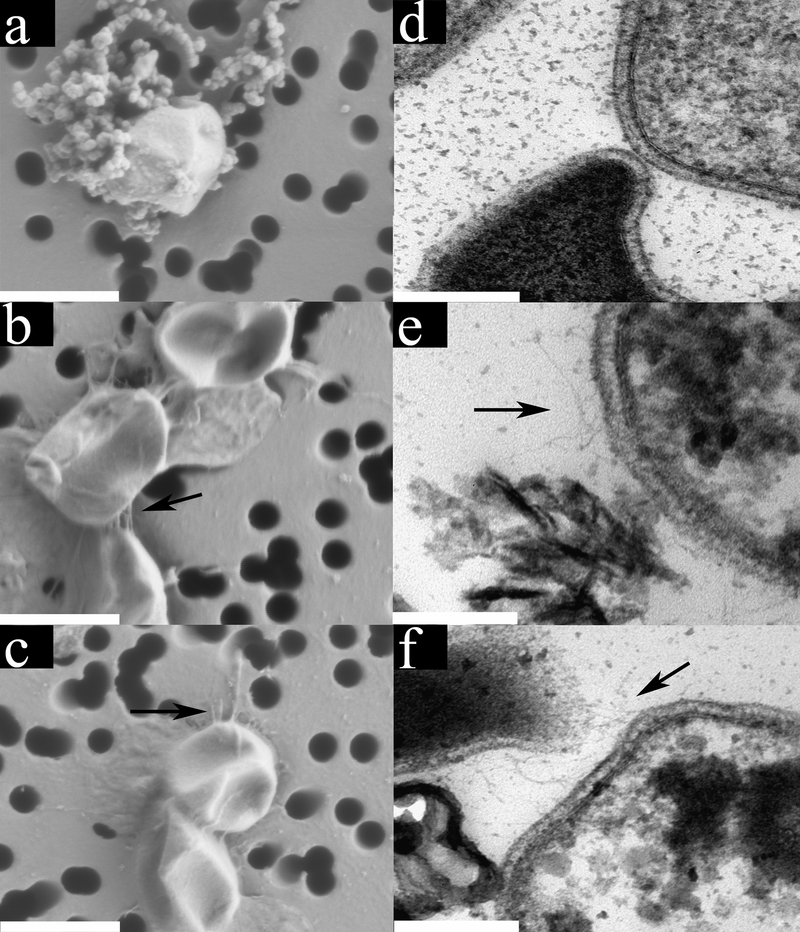
Fig. 1. Electron micrographs of DS80 cells grown with different electron donor/acceptor pairs reveal differences in morphology.
Field emission scanning electron micrographs (FE SEMs) of H2/S° (a), S°/Fe3+ (b), and H2/Fe3+ (c) grown cells with arrows denoting pili-like structures where present. Globules outside cells in panel a correspond to nanoparticles of S° 20. FE SEM scale bars represent 1000 nm. Thin section transmission electron micrographs (TEMs) of cells grown with H2/S° (d), S°/Fe3+ (e), and H2/Fe3+ (f) with arrows denoting pilin-like structures where present. Insoluble precipitates of unknown composition are visible in panel e and f. TEM scale bars represent 200 nm.

