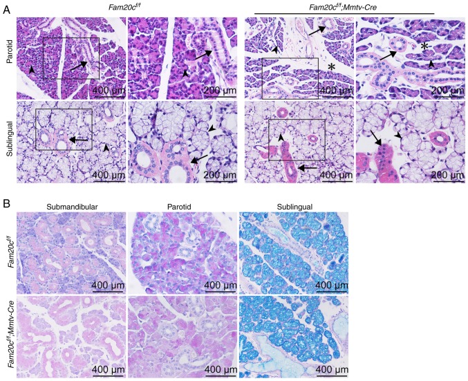
Figure 3.
PG/SLG morphological and structural alterations in cKO mice. (A) Hematoxylin and eosin staining of the PG and SLG. There was more mesenchymal tissue (indicated by asterisks) present within the mutant PG. The number of ducts (arrows) and acinar cells (arrowheads) did not change substantially. (B) Periodic Acid-Schiff staining of the SMG, PG and SLG. The production of mucin by the SMG did not change appreciably between the cKO and control mice. Fam20C, family with sequence similarity 20-member C; PG, parotid gland; SLG, sublingual gland; SMG, submandibular gland; cKO, Fam20cf/f; Mmtv-Cre conditional knockout.

