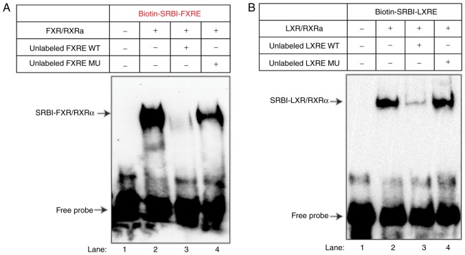
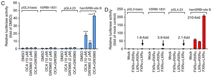
Figure 4.
EMSA and reporter analyses of the association of FXR and LXR with the regulatory intronic region of site B segment of the hamster SR-BI gene. (A) EMSA to evaluate DNA-binding by FXRα/RXRα. A biotin-5′ end-labeled SRBI-FXRE probe was incubated without (lane 1) or with 100 ng of FXRα and 100 ng of RXRα recombinant proteins in the absence (lane 2) or presence of 100-fold molar excess of unlabeled WT probe (lane 3) or MU probe (lane 4). The protein-DNA complex was resolved on 5% TBE gels, transferred onto a nylon membrane and UV-crosslinked to the membrane prior to DNA visualization by avidin-chemiluminescent probe. The data shown are representative of three separate EMSA assays with comparable results. (B) EMSA to evaluate DNA-binding by LXRα/RXRα. Biotin-5′ end labeled SRBI-LXRE probe was incubated without (lane 1) or with 100 ng of LXRα and 100 ng of RXRα recombinant proteins in the absence (lane 2) or presence of 100-fold molar excess of unlabeled WT probe (lane 3) or MU probe (lane 4). The protein-DNA complex was resolved on 5% TBE gels, transferred to a nylon membrane and UV-crosslinked prior to DNA visualization by avidin-chemiluminescent probe. The data shown are representative of two separate EMSA assays with comparable results. (C) HepG2 cells were cotransfected with pGL3-basic, pGL3-hSRBI-1831, pGL4.23, or hamster site B WT plasmid and indicated nuclear receptor expression plasmids or the control vector (pCMV-empty). At 2 days post-transfection, cell lysates were prepared. Following normalization with Renilla activity, the relative luciferase activity (fold of mock) is indicated for each reporter plasmid. (D) Human and hamster SRBI reporter vectors and their respective control vectors were transfected into HepG2 cells along with pRL-TK. The following day, cells were cultured in MEM containing 0.5% fetal bovine serum overnight and treated with OCA (10 µM), GW3965 (2 µM), or GW3965 + OCA for 24 h prior to cell lysis. Data are presented as the mean ± standard error of the mean of four replicates per treatment and are expressed as ratio of firefly/Renilla activity from each sample where the relative luminescence from DMSO-treated cells is set to 1. Statistical significance among all groups was assessed by one-way analysis of variance with Tukey's multiple comparison test. **P<0.01 and ***P<0.001 compared with DMSO-treated samples. The data shown are representative of three separate transfection experiments. FXR, farnesoid X receptor; FXRE, FXR response element; LXR, liver X receptor; LXRE, LXR response element; SR-BI, scavenger receptor class B type I; WT, wild-type; mu, mutant; OCA, obeticholic acid.


