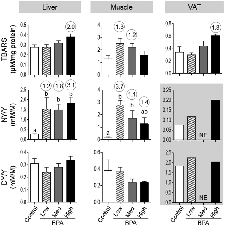
Figure 3:
Mean ± SEM tissue TBARS and oxidized tyrosine concentrations in control, BPAlow, BPAmed and BPAhigh dose prenatal BPA-treated groups in the liver (left), muscle (middle) and VAT (right panel). Numerical superscripts are Cohen’s d values ≥ 0.8 comparing control and different dose groups of BPA. Mean values of oxidized tyrosine concentrations in VAT that is previously published [21] are shown in the grey area (NE = Not Examined). Differing letters in histograms (NY/Y in liver and muscle) indicate statistically significant differences; a vs. b: p<0.05; ab not different from a or b.

