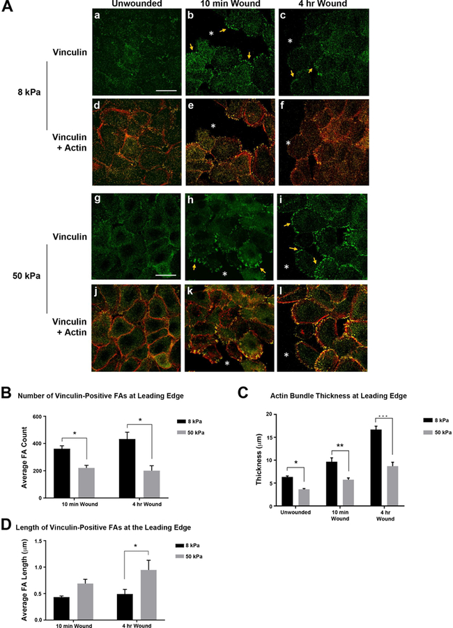
Figure 4. Increased surface stiffness reduces focal adhesion number and actin bundle thickness along the leading edge.

Human corneal limbal epithelial cells were cultured on fibronectin-coated 8 kPa and 50 kPa substrates until confluent. Scratch wounds were made and cultures were fixed 10 minutes and 4 hours after injury. Cells were stained for vinculin and counter stained with rhodamine phalloidin to detect F-actin. Images were obtained using a 63x objective on a Zeiss Axiovert LSM 700 confocal microscope. Staining with the secondary antibody was performed to set the negative level and all experimental conditions were imaged at the same setting as was used for secondary antibody alone. Images represent a minimum of 4 independent experiments. A.a,g. Localization of vinculin in unwounded cells is affected by substrate stiffness 8 or 50 kPa. A.d,j. Vinculin and F-actin (red). A. b-c,g-i. Vinculin is localized within focal adhesions after injury (arrows). A. e-f,k-l. Vinculin and F-actin (red). *wound edge. Scale bar is 25 µm. B-D. Leading edges were analyzed in 500 µm intervals. Actin bundle thickness along with focal adhesion number and length were determined using FIJI/ImageJ (NIH, Bethesda, MD; http://imagej.nih.gov/ij/). Statistical analysis of each time point and condition was conducted (Two way ANOVA). B. Decreased number of vinculin-positive focal adhesions on stiffer substrates. C. Actin bundle thickness at the leading edge is reduced in cells on stiffer substrates for all conditions. Thickest regions of actin were measured in cells along the wound edge. D. Vinculin focal adhesion length increases with increased substrate stiffness 4 hr after injury. Standard error bars are ± S.E.M. *P < 0.05, **P < 0.01, ***P < 0.005.
