
Figure 5. Vinculin pY1065 localization and phosphorylation are affected by increased substrate stiffness.
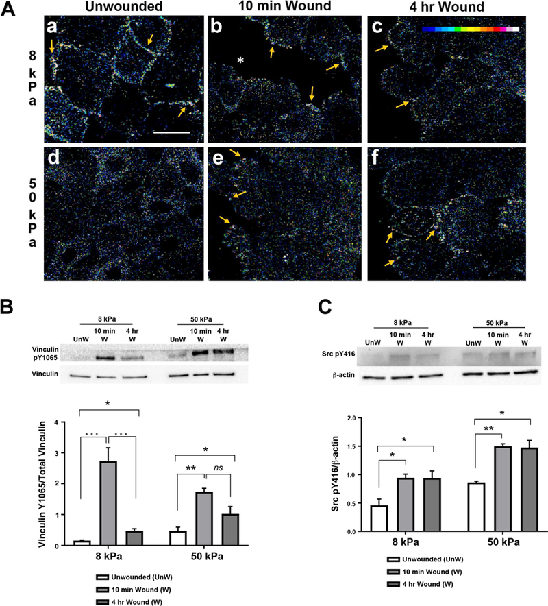
HCLE cells were cultured and scratch wounds made. Immunostaining is shown as changes in fluorescence intensity. Images were obtained using a 63x objective on a Zeiss Axiovert LSM 700 confocal microscope. Secondary antibody controls were set to negligible levels and all experimental cell conditions were imaged at same setting that was used for secondary antibody alone. A. In unwounded cultures, vinculin pY1065 localizes to membranes on 8 kPa substrates but remains diffuse throughout cells on 50 kPa substrates. Arrows indicate vinculin pY1065 at the leading edge and on cell membranes. *, wound edge. Scale bar is 25 µm. B-C. Western blot analysis was conducted on samples 10 minutes and 4 hours after injury. Protein was extracted, resolved by 10% SDS-PAGE, and immunoblotted for vinculin pY1065 and Src pY416. Relative expression of vinculin pY1065/total vinculin and Src pY416/ β-actin was determined by quantifying band intensity. Representative blots are shown. Unw, unwounded. B. Phosphorylation of tyrosine 1065 on vinculin remains elevated after injury in cells on stiffer substrates. C. Src phosphorylation remains elevated after injury on 8 kPa and 50 kPa substrates. Data represent a minimum of 4 independent experiments. Standard error bars are ± S.E.M. Statistical analysis conducted using Two way ANOVA. *P < 0.05, **P < 0.01, ***P < 0.005.