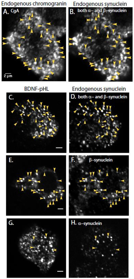
Fig. 1. Endogenous synuclein labels secretory granules in non-transfected cells (A, B) and newly synthesized chromaffin granules expressing exogenous BDNF-pHl in transfected cells (C-H).
Granules were identified with an antibody against chromogranin A (A) and endogenous synuclein with a monoclonal antibody that recognizes both the α- and β-isoforms of bovine synuclein (DSHB H3C, B). Immunocytochemistry was performed on chromaffin cells transiently expressing the chromaffin granule marker BDNF-pHl (C-H). (D) Endogenous synuclein was again detected by DSHB H3C, which recognizes both α- and β-synuclein. Arrowheads indicate some of the co-localized puncta. Synuclein puncta in a neighboring non-transfected cell are visible at the bottom of the image. (F) Endogenous synuclein as detected by an antibody specific for β-synuclein (Abcam 15532); (H) Endogenous synuclein as detected by an antibody specific for α-synuclein (Abcam 138501). Panels A and B, C and D, E and F and G and H are paired images of the same field of view. Note that panels C, E, and G show single transfected cells. Panels D, F, and H show several cells including the transfected cell. Scale bars = 2 μm.

