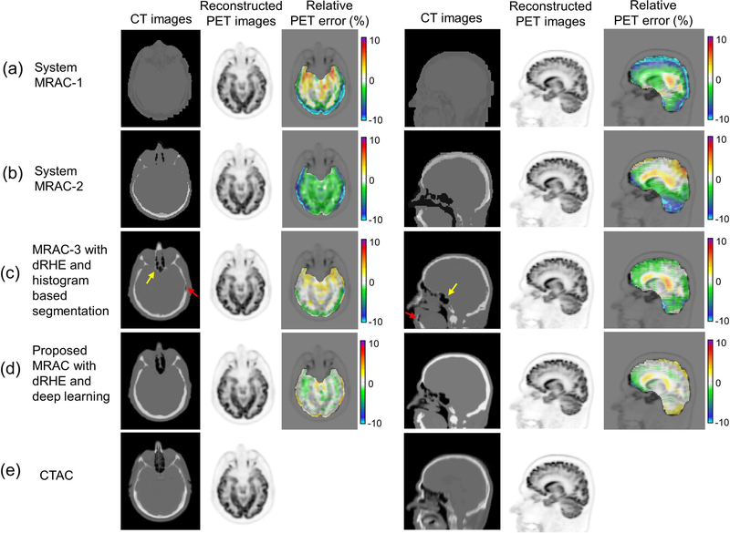
Fig. 4.
PET reconstruction. An axial slice and a coronal slice in a CT images, a reconstructed PET image, and a relative PET error map in (a) MRAC-1, (b) MRAC-2, (c) MRAC-3, the proposed MRAC with deep learning, and (e) CTAC. As seen in PET error maps the proposed MRAC shows much reduced percent error (< 1%) in most brain regions over other MRACs.

