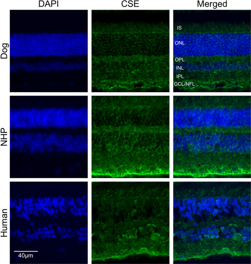
Figure 6.
Immunohistochemical analysis of CSE labeling in normal retina from canine, non-human primate and human. Retinal cryosections from (A-C) canine, non-human primate (D-F) and human (G-I). Immunolabeling showed RPE layer, and mainly in GCL. DAPI was used to stain nuclei. (Scale bar: 20 µm; GCL: ganglion cell layer; NFL: nerve fiber layer; IS: Inner segment layer of photoreceptor cells).

