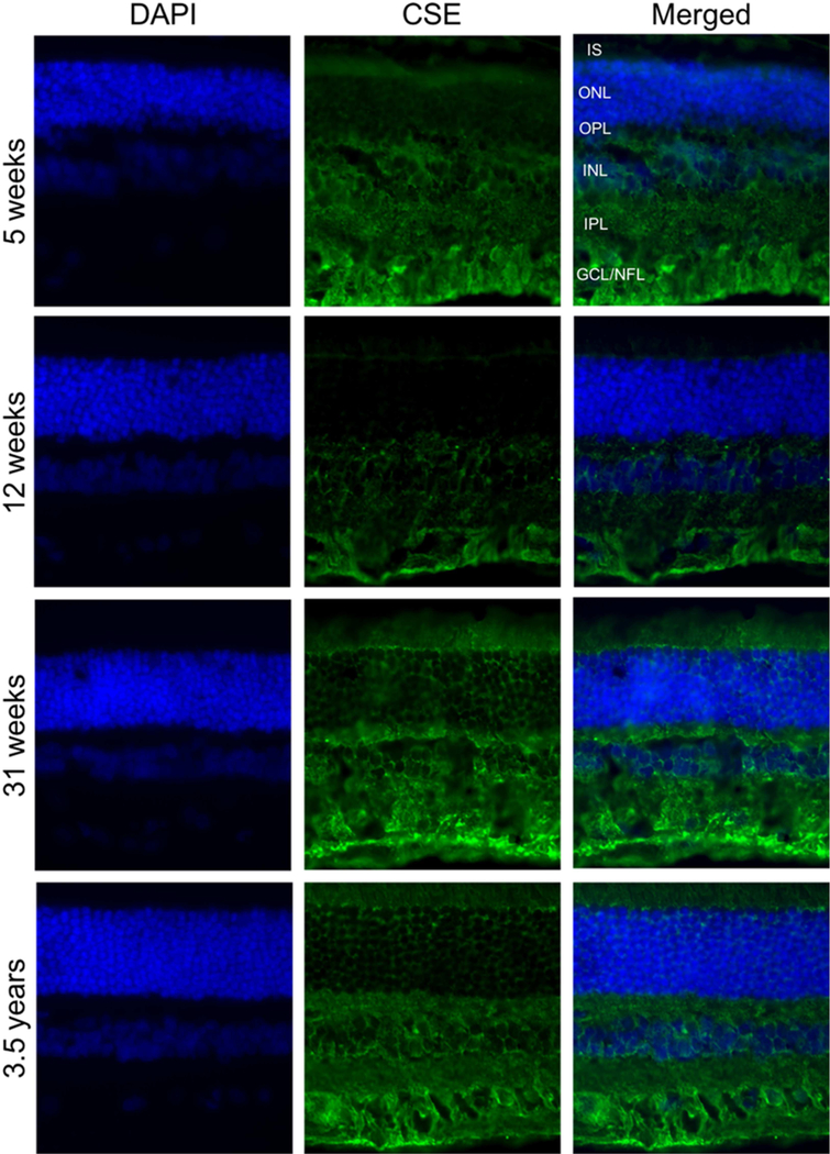
Figure 7.
Immunohistochemical analysis of CSE in canine retinas at different ages. Retinal cryosections from (A, B, C) five weeks, (D, E, F) twelve weeks, (G, H, I) thirty-one weeks and (J, K, L) three and a half years old dogs were stained with antibody against CSE. CSE (green fluorescence) was mainly in GCL. DAPI was used to stain nuclei (A, D, G and J). C, F, I and L are merged pictures. (Scale bar: 20 µm; PR: Photoreceptor cells, GCL: ganglion cell layer; NFL: nerve fiber layer; IS: Inner segment layer of photoreceptor cells).

